The word “autopsy” comes from the roots autos (“self”) and opsis (a sight, or seeing with one’s own eyes)- so an autopsy is the examination of a body after death by someone of like species- another human.
So what do you call the post-mortem examination of an animal? The appropriate term is “necropsy,” derived from necro (“death”) and the aforementioned opsis.
So, all autopsies are necropsies, but not all necropsies are autopsies! In both instances, the procedure is the dissection of a body to determine why the individual died. Here at Cornell Wildlife Health Lab, we perform necropsies to determine the cause of death so that we can keep an eye out for diseases and toxins that can impact wildlife, domestic animals or humans.

Before the physical examination, we learn as much as we can about the history of the animal. Since we work exclusively with wildlife, this information is typically scarce and limited to location, environmental conditions, and if the animal was alive or dead when it was found.
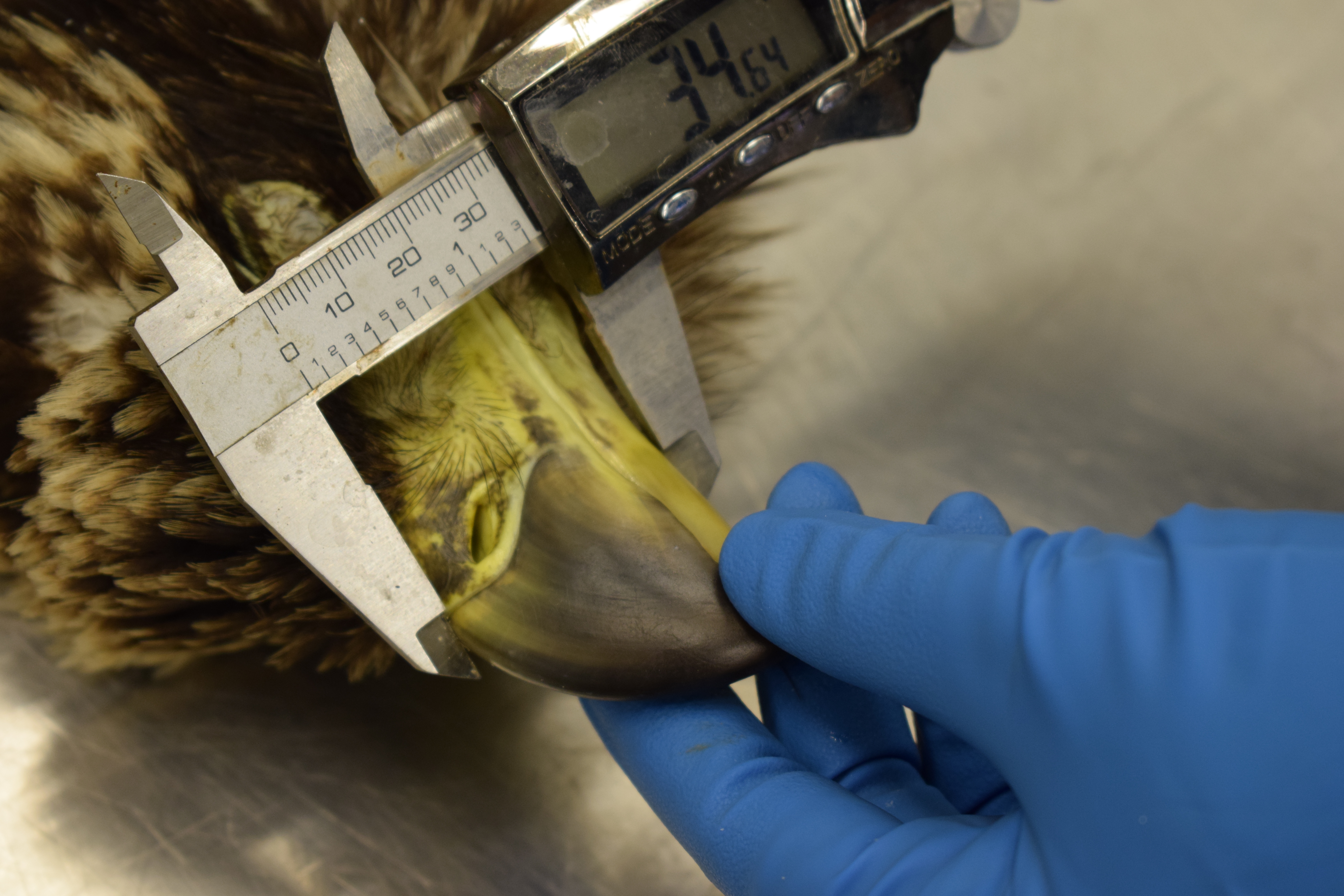
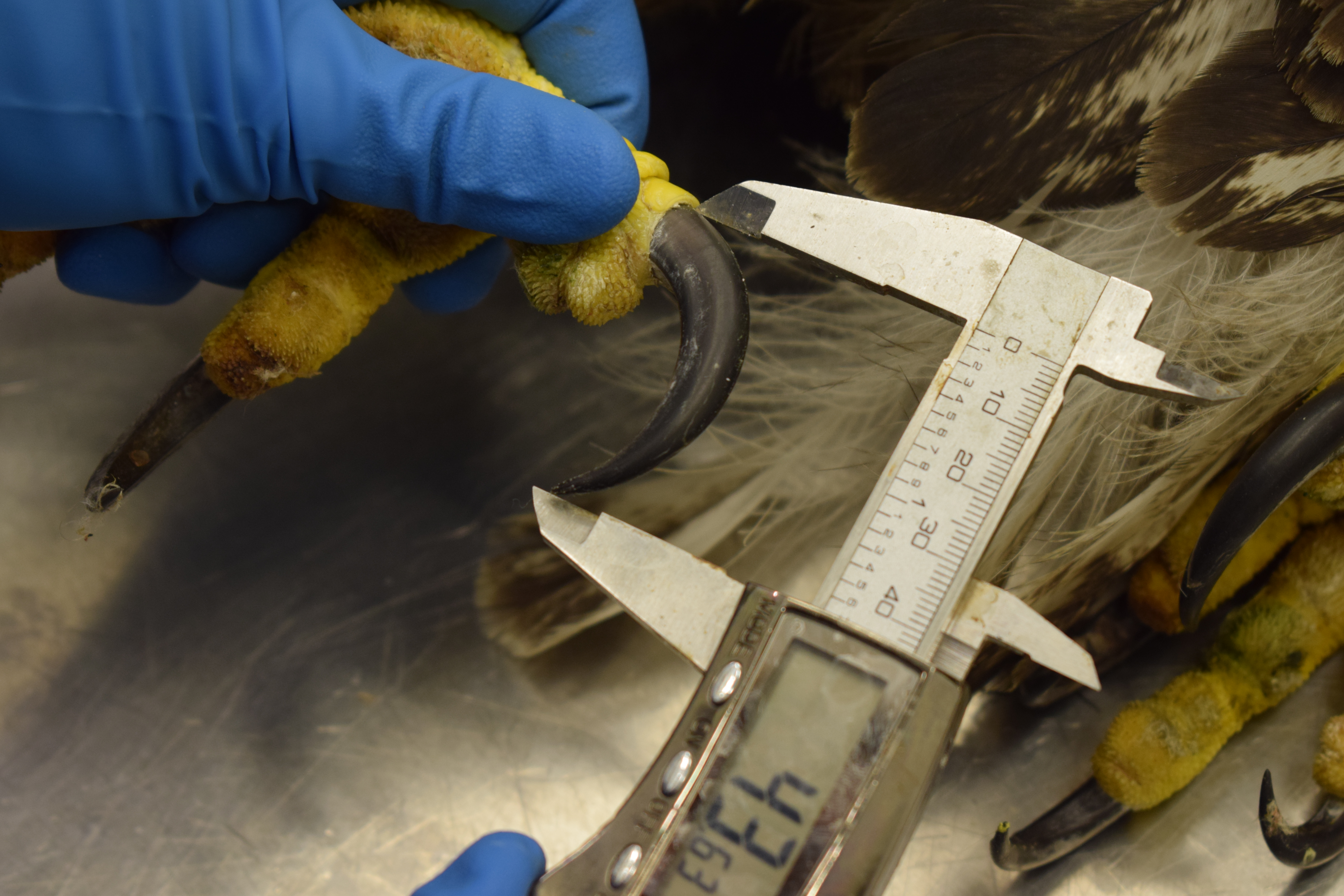
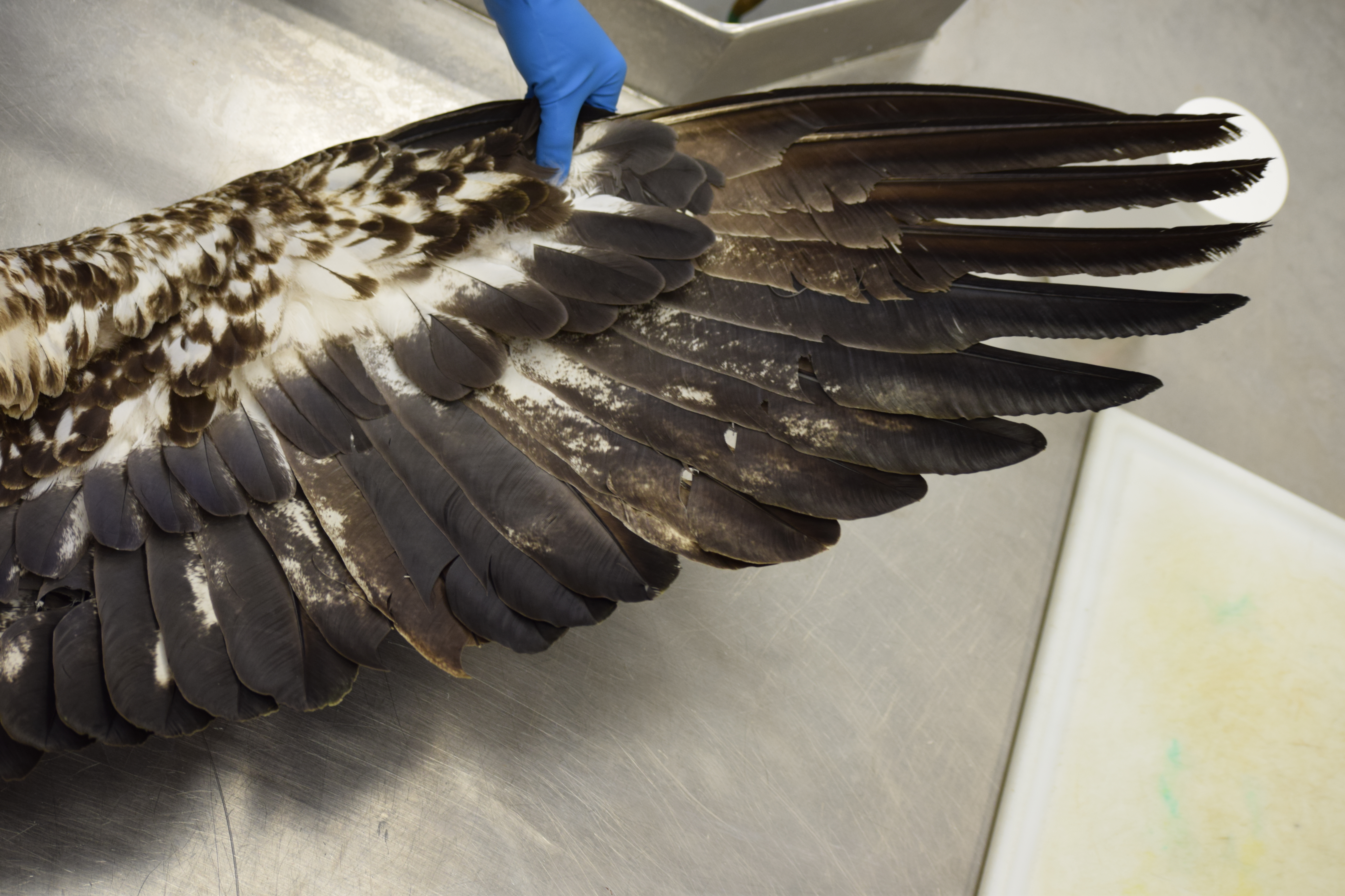
From here, a veterinary pathologist, (a specialist in diagnosing disease by examining animal tissues), will attend to the case. Special facilities and personal protective gear like masks and gloves are used to protect the pathologist from any disease that is transferable between animals and humans, like rabies or West Nile Virus (called a zoonosis). They record any identifying marks like a notch code on a turtle shell or leg band/wing tags on a bird, as well as weight, sex and age if it can be determined from feathers or teeth. It is important to note any visible abnormalities, which could range from fur loss, to tumors, to bullet wounds, and everything in between.
Click on images to view them larger
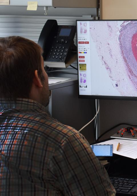
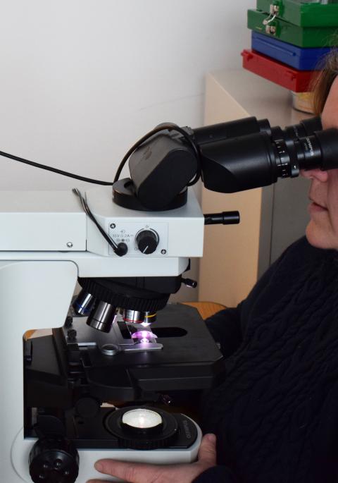
An internal examination follows the external exam. All organs are first looked at "in situ"- in place within the body cavity- to evaluate position, size, and color. This can be made difficult if the animal is autolyzed (too decomposed), or has been scavenged. Each organ is then removed for examination and sampling. For straightforward cases like trauma from being hit by a car, the process ends here. For others we collect samples in duplicate, fixed and fresh. Fixed tissues are preserved in formalin for histopathology, the microscopic examination of the tissues. Fresh samples are sent for testing such as bacteriology, molecular diagnostics, parasitology, toxicology, or virology. Some samples are saved for use in future research projects. Other samples are collected for the tissue archive to aide researchers.

Once we have all the information from testing, microscopic exam, and history, we make a diagnosis of the cause of death. We can't always get an answer on every case, but with over 9,000 cases so far, we have a fair amount of information on what impacts wildlife in NY. We use that to identify and work on issues that cause harm, as well as pick up new introduced diseases before they can get too far. Necropsy is just one tool that we have at our disposal to increase our knowledge of wildlife disease.