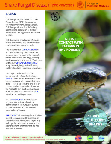
What is Snake Fungal Disease?
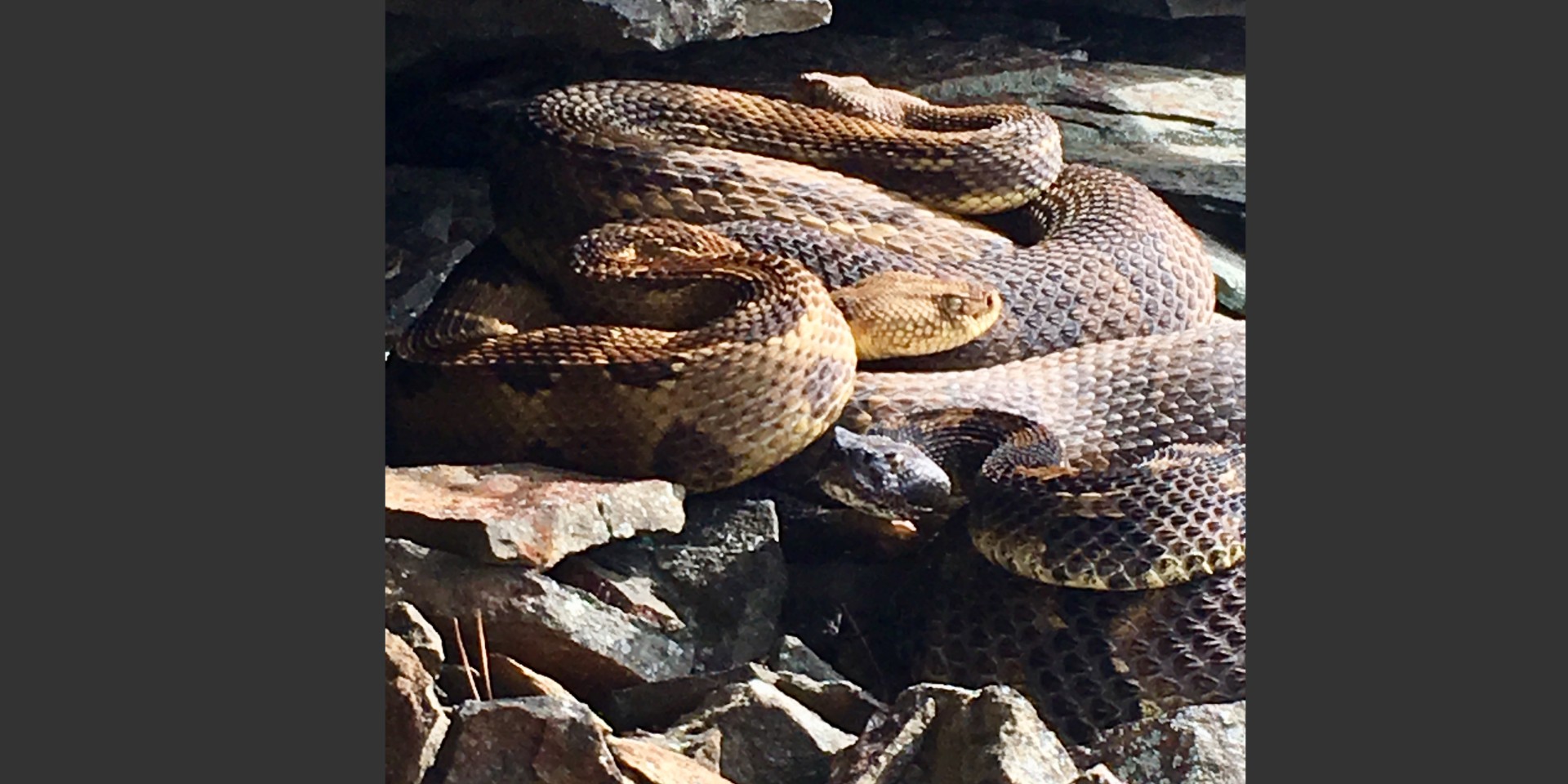
Snake Fungal Disease (SFD) is caused by the fungus Ophidiomyces ophiodiicola and it poses a significant threat to wild snakes in the eastern United States. First discovered in 2006 in a declining New Hampshire population of timber rattlesnakes (Crotalus horridus), SFD has now been recorded in over a dozen species. Like other recently emerged fungal diseases of wildlife, snake fungal disease is often fatal and difficult to control in the environment. SFD is an emerging infectious disease with serious conservation implications for native snake populations, particularly rattlesnakes. Habitat loss, climate change, and human persecution have caused fragmentation of populations of many native snake species (particularly rattlesnakes). Snake fungal disease now has the capacity to drive these last remaining populations to extinction.
SFD_TRattlesnake MF.jpg
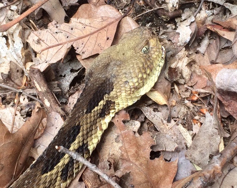
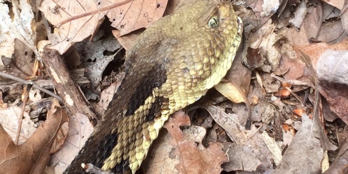
Timber rattlesnake with SFD starting on the mouth, photo by Melissa Fadden
It was 2016 when research determined O. ophiodiicola to be the main cause of SFD. Transmission is by direct contact with infected animals or a contaminated environment. A few days following exposure, snakes develop discolored skin at the site of infection. As the infection progresses, fungus penetrates the tough outer layer of the skin and creates inflamed, crusted lesions. If the fungus remains contained in the skin, the disease can sometimes resolve when a snake sheds. However, some fungal hyphae (stalks) may remain behind and cause reinfection.
Despite the ability for some snakes to naturally resolve their infection, the disease can cause physiological and behavioral changes that reduce survival. Some facial lesions may make catching prey and swallowing difficult and painful. When the animal is weakened and unable to mount a good immune response, fungal infections can become severe.
Can you treat SFD?
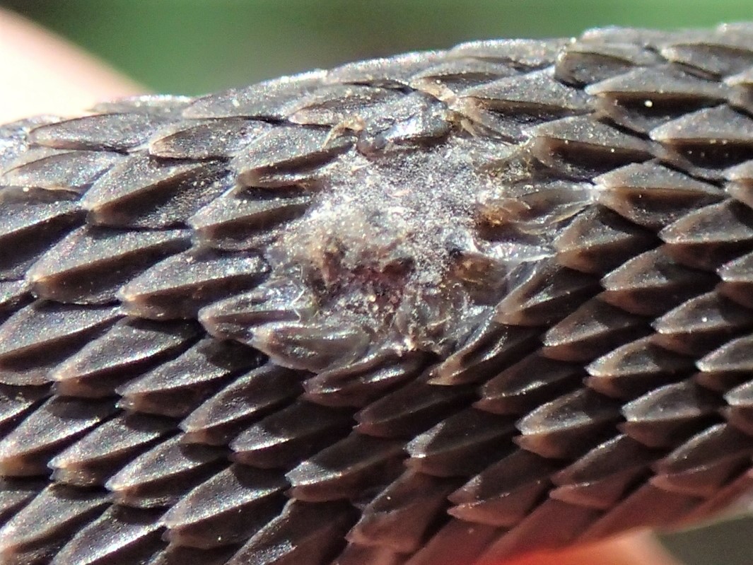
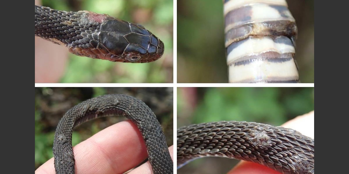
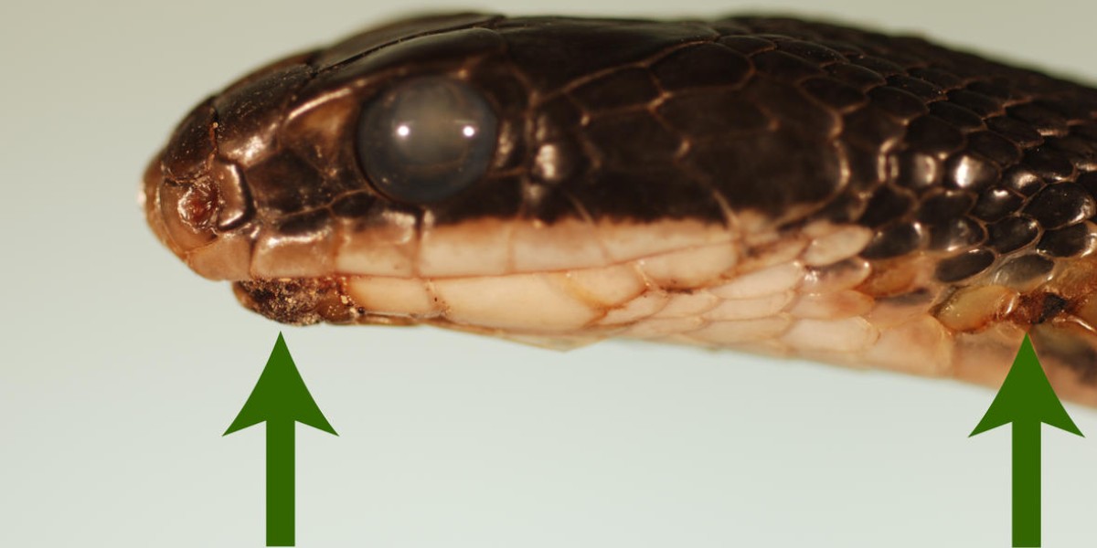
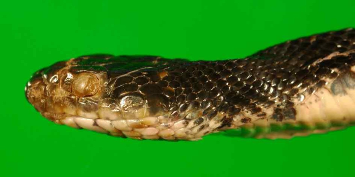
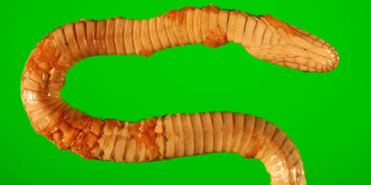
Clinical signs associated with SFD vary depending on the stage and severity of the disease. Generally, most infected snakes have localized scabs and crusty, thickened skin lesions. Other common clinical signs include blisters, swelling, cloudy/opaque eyes and lumps under the skin. In more severe forms of the disease, disfiguring facial lesions can develop.
SFD web close up.jpg
In the laboratory, SFD can be diagnosed using culture, PCR (DNA based test) and histopathology. Skin lesions may need to be treated surgically, and thermal/fluid/nutritional support and the use of both systemic and topical antifungals can help.
Where is Snake Fungal Disease found?
Snake fungal disease was initially described and diagnosed in 2006 in a New Hampshire population of timber rattlesnakes. By 2007, this population had suffered a 50% decline due to the disease. In 2008, the disease appeared in populations of eastern massasauga rattlesnakes in Illinois, and this species is at the brink of extinction in the state. The disease has been reported in wild snakes over much of the eastern United States.
- Vermont
- New York
- Massachusetts
- Connecticut
- New Jersey
- Pennsylvania
- Virginia
- Ohio
- Kentucky
- Tennessee
- South Carolina
- Georgia
- Alabama
- Louisiana
- Wisconsin
- Minnesota
- Missouri
- Eastern foxsnake (Pantherophis vulpinus)
- Ring-necked snake (Diadophis punctatus)
- Northern copperhead (Agkistrodon contortrix)
- Northern pinesnake (Pituophis melanoleucus)
- Cottonmouth (Agkistrodon piscivorus)
- Northern watersnake (Nerodia sipedon)
- Eastern racer (Coluber constrictor)
- Eastern ratsnake (Pantherophis obsoletus)
- Timber rattlesnake (Crotalus horridus)
- Eastern massasauga (Sistrurus catenatus)
- Pygmy rattlesnake (Sistrurus miliarius)
- Plains gartersnake (Thamnophis radix)
- Eastern gartersnake (Thamnophis sirtalis sirtalis)
- Mudsnake (Farancia abacura)
- Milksnake (Lampropeltis triangulum)
SFD is Not the Only Infectious Fungal Disease Impacting Wildlife
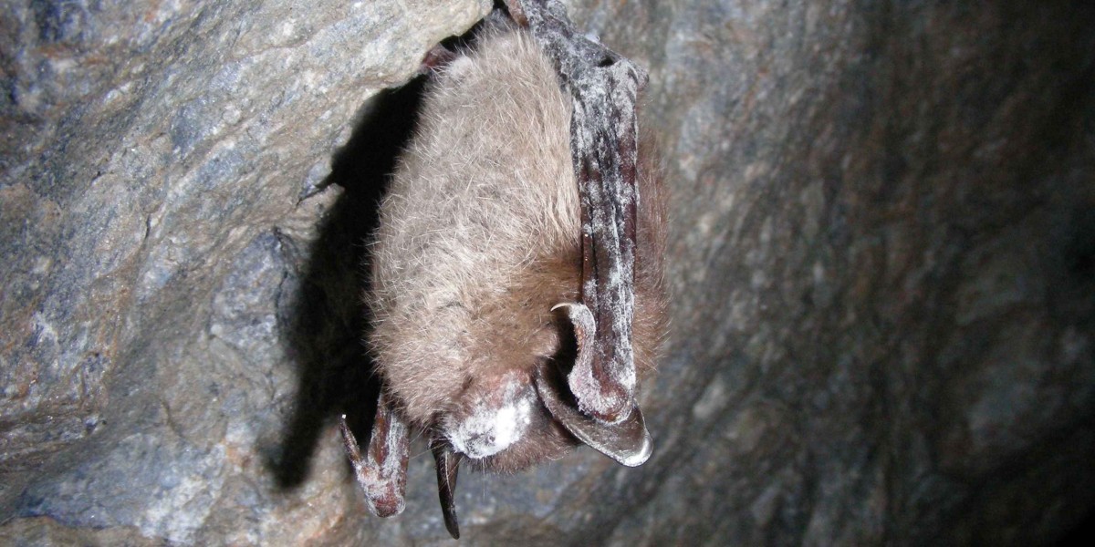
In the past few decades several infectious fungal diseases of wildlife have caused widespread population declines and sometimes extinction of species. In addition to SFD, these include white-nose syndrome (North American bats), and chytridiomycosis in amphibians. White-nose syndrome is caused by the fungus Pseudogymnoascus destructans and has caused the deaths of millions of bats in the northeastern United States since its emergence in 2006. Chytridiomycosis is a skin disease caused by either of two species of chytrid fungus, Batrachochytrium dendrobatidis (Bd) and Batrachochytrium salamandrivorans (Bsal). Since its discovery as a pathogen in the 1990s, Bd has caused the decline or extinction of approximately 200 Anura (frog) species, representing the most significant extinction event in modern history. The recently discovered Bsal is threatening salamander populations in Europe, particularly the fire salamander (Salamandra salamandra), which has suffered population declines of over 99% in the Netherlands. Together, these diseases pose a significant and imminent threat to global biodiversity.


![Slimy salamander (Plethodon glutinosus) leaf in Land Between the Lakes, By AugustTulipa13 (Own work) [CC BY-SA 4.0 ] via Wikimedia Commons Slimy salamander (Plethodon glutinosus) leaf in Land Between the Lakes](https://cwhl.vet.cornell.edu/system/files/styles/slideshow/private/media/Slimy_salamander_%28Plethodon_glutinosus%29_leaf_in_Land_Between_the_Lakes_0.jpg?itok=PSGtCTkQ)
