Neospora is a cyst-forming coccidian protozoan parasite (Neospora caninum) which is in the family Sarcocystidae. It is structurally and biologically similar to Toxoplasma gondii and, until 1988, was commonly misdiagnosed as such.
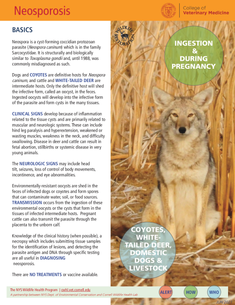
Dogs and coyotes are definitive hosts for Neospora caninum, and cattle and white-tailed deer are intermediate hosts. Only the definitive host will shed the infective form, called an oocyst, in the feces. Ingested oocysts will develop into the infective form of the parasite and form cysts in the many tissues.
Clinical signs develop because of inflammation related to the tissue cysts and are primarily related to muscular and neurologic systems. These can include hind leg paralysis and hyperextension, weakened or wasting muscles, weakness in the neck, and difficulty swallowing. Disease in deer and cattle can result in fetal abortion, stillbirths, or systemic disease in very young animals. The neurologic signs may include head tilt, seizures, loss of control of body movements, incontinence, and eye abnormalities.
Environmentally-resistant oocysts are shed in the feces of infected dogs or coyotes and form spores that can contaminate water, soil, or food sources. Transmission occurs from the ingestion of these environmental oocysts or the cysts that form in the tissues of infected intermediate hosts. Pregnant cattle can also transmit the parasite through the placenta to the unborn calf.
Knowledge of the clinical history (when possible), a necropsy, which includes submitting tissue samples for the identification of lesions, and detecting the parasite antigen and DNA through specific testing are all useful in diagnosing neosporosis.
There are no treatments or vaccines available.
Coyotes and domestic dogs have been confirmed as definitive hosts and, because of their similar diets and close relationship to these other canids, it is very likely that gray wolves can serve as definitive hosts as well. Research has been ongoing to determine other carnivores that can serve as definitive hosts and antibodies have been detected in other wolf species, as well as multiple fox species and raccoons.
White-tailed deer have been confirmed in recent years to be natural intermediate hosts and many other species of herbivores have tested positive for antibodies to N. caninum, indicating they could serve as intermediate hosts. These include moose, bison, caribou, and musk ox. Neospora caninum has also been identified in brown rats and antibodies have been detected in seven species of marine mammals.
The oocysts containing the zygote stage of N. caninum are passed in the feces of the definitive host (dog or coyotes) and then produce spores once in the environment. Once ingested, they then become tachyzoites or bradyzoites which are both asexually reproductive stages of N. caninum.
Tachyzoites rapidly reproduce in the cells and may travel through the placenta of a pregnant cow to infect an unborn calf. Bradyzoites reproduce slowly and form cysts in the tissues of the intermediate hosts. These are then consumed by a definitive host feeding on the tissues and reproduce sexually in the definitive host to form the oocysts.
Laboratory testing to diagnose Neospora infection includes immunohistochemistry, serology, PCR, genotyping, and isolation. Histopathology can be used to identify parasites and lesions in the brain, spinal cord, heart, skeletal muscle, liver, and placenta.
Separation of domestic and wild animals may help to reduce the transmission between domestic dogs, cattle, and wildlife.