Echinococcosis, also known as Cystic Hydatid Disease, is an infection of tapeworm larvae of Echinococcus granulosus, a parasitic cestode with a multi-host lifecycle.
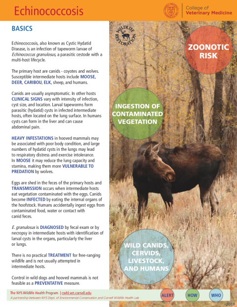
The primary hosts are canids - coyotes and wolves. Susceptible intermediate hosts include moose, deer, caribou, elk, sheep, and humans.
Canids are usually asymptomatic. In other hosts clinical signs vary with intensity of infection, cyst size, and location. Larval tapeworms form parasitic (hydatid) cysts in infected intermediate hosts, often located on the lung surface. In humans, cysts can form in the liver and can cause abdominal pain.
Heavy infestations in hooved mammals may be associated with poor body condition, and large numbers of hydatid cysts in the lungs may lead to respiratory distress and exercise intolerance. In moose, it may reduce the lung capacity and stamina, making them more vulnerable to predation by wolves.
Eggs are shed in the feces of the primary hosts, and transmission occurs when intermediate hosts eat vegetation contaminated with the eggs. Canids become infected by eating the internal organs of the hoofstock. Humans accidentally ingest eggs from contaminated food, water or contact with canid feces.
E. granulosus is diagnosed by fecal exam or by necropsy in intermediate hosts with identification of larval cysts in the organs, particularly the liver or lungs.
There is no practical treatment for free-ranging wildlife, and it is not usually attempted in intermediate hosts.
Control in wild dogs and hooved mammals is not feasible as a preventative measure.
Two distinct forms of E. granulosus are recognized in North America: a northern type found in forest habitats with a wildlife transmission cycle involving free-ranging wolves and deer, and a southern European type with a domestic animal life cycle involving hooved animals and dogs.
Adult tapeworms, typically 3-6mm long, reside in the small intestine of carnivorous definitive hosts – namely dogs, coyotes or wolves (canines). Reproductive adults lay eggs in the small intestine that pass in the animal’s feces. Although vulnerable to heat and drying out, E. granulosus eggs are highly resistant to environmental stressors, including freezing. They can survive for at least a year, particularly in damp and cool conditions.
Intermediate hosts may include moose, elk, caribou, or deer in sylvatic cycles and sheep, goats, cattle, or pigs in pastoral cycles.
Reproductive adults in the small intestine of canine hosts lay eggs that are passed in the animal’s feces. Each worm may produce up to 1000 eggs every 10 days for up to 2 years. Once in the environment, eggs may be transported by wind, water, or insects to contaminate vegetation or water sources.
Within the intermediate host, the eggs hatch in the small intestine and release hooked embryos that penetrate the intestinal wall, enter the bloodstream, and travel primarily to the lungs or, occasionally, to the liver, kidneys, brain or bone marrow. In the lung, the larvae form a single-chambered, fluid-filled hydatid cyst ranging from 2 to 30 cm in diameter. Within the cyst, thousands of larval tapeworms or protoscolices (essentially tiny tapeworm heads) are produced by an asexual budding process.
These protoscolices are infective to the definitive canine hosts, which become infected when they consume the cyst-containing organs of intermediate hosts. In sylvatic cycles, this occurs when wolves and coyotes prey upon or scavenge an infected animal; in pastoral cycles, this often occurs due to the practice of feeding viscera from home-slaughtered sheep to domestic dogs. Once ingested by the canine, the protoscolices attach to the wall of the small intestine and mature into adult worms, which lay eggs to complete the life cycle.
Although not a normal part of the tapeworm’s life cycle, humans may be infected by ingestion of eggs in contaminated food or water or by hand-to-mouth transmission after handling infected canids or their fecal material. In humans, the eggs hatch in the gut, and larvae travel via the bloodstream to the liver, lungs, brain, skeletal muscle, or eye, with the liver being the most common destination.
In humans, discomfort, abdominal pain, nausea, and vomiting have been reported when liver cysts grow large enough to place pressure on surrounding organs. Cyst rupture and subsequent release of cystic fluid may cause mild to severe anaphylactic (allergic) reactions.
Canine definitive hosts may be treated with praziquantel, an anti-parasitic drug. In humans, infections are typically treated by surgical removal of the hydatid cysts paired with drug therapy to kill any tapeworm larvae that may remain in the body. In some cases, chemotherapy, cyst puncture, or percutaneous aspiration, injection of chemicals, and reaspiration (PAIR) may be used as alternatives to surgery.
In the canine definitive hosts, diagnosis is made by identifying adult worms in the feces or small intestine, or by testing feces for E. granulosus antigens by ELISA. Serological testing may also be used to detect antibodies to E. granulosus antigens but will not distinguish between current infections and previous exposure.
In humans, infection is diagnosed by ELISA or serological testing. Cysts may be detectable by imaging techniques such as CT scans, ultrasound, or MRI, although additional testing would be necessary to confirm the diagnosis.
To prevent infection, humans should wear gloves when handling canine feces and wash their hands after handling feces and before eating. Domestic dogs should be dewormed regularly and should not be fed uncooked meat from deer, elk, moose or sheep, or allowed to scavenge.
Control in livestock may be accomplished by vaccination with a protein from the parasite’s egg; however, this vaccine has not been effective in preventing infection of deer.