

Image
Rabies
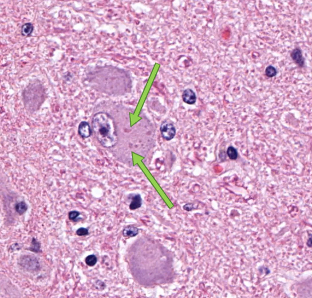
One of the most frequent questions the public or the biologists have when they find a mammal acting abnormally or dead is “does it have rabies?” We send all of our samples to the NY State Department of Health (DOH) for official rabies testing, but we sometimes will look at histology of the brain of suspected rabid animals under the microscope, particularly if the sample that we sent to the DOH was not ideal. In the case pictured, the rabies test at DOH was positive. Numerous rabies virus inclusion bodies are present in the cytoplasm of the large cell in the middle of the picture. These are called Negri bodies and are diagnostic for rabies virus infection.